

Preview
Creation Date
9-28-2017
Keywords
Magnesium oxalate; Diamond Anvil Cell; Optical microscope; Irradiation; Hard x-rays
Disciplines
Arts and Humanities | Life Sciences | Physical Sciences and Mathematics | Social and Behavioral Sciences
Document Type
Image
File Format
jpeg
File Size
542 Kb
Language
English
Description
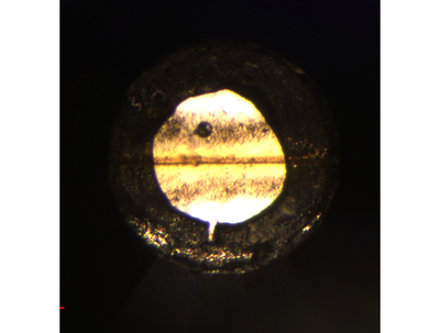
A sample of magnesium oxalate, confined within a 100 μm diameter beryllium gasket, pressurized inside of a Diamond Anvil Cell to about 100,000 times atmospheric pressure. The picture was taken with an optical microscope looking through a diamond. The sample was irradiated with 10 keV x-rays produced at the Advanced Photon Source at the Argon National Lab in Chicago. The yellow area shows material that has been decomposed to a doped form of polymeric carbon monoxide by exposure to hard x-rays, while white area is unadulterated Magnesium Oxalate.
Time Period
2010-2019; 21st century
Keywords
Magnesium oxalate; Diamond Anvil Cell; Optical microscope; Irradiation; Hard x-rays